What to Expect During an Eye Exam

The parts of a comprehensive eye examination vary according to the patient's age, date of last exam, and other factors. Not all parts of the eye exam may be needed or performed, but the first part of the eye exam will include documenting medical history. Here are some eye and vision tests that are likely to be encountered during a comprehensive eye exam:
Visual Acuity Tests
Visual acuity tests measure the sharpness of vision and are usually performed using a projected eye chart to measure the distance visual acuity and a hand-held small acuity chart to measure the near vision (for reading).
Color Blindness Test
A screening test that checks the color vision is often performed early in a comprehensive eye exam to rule out color blindness.
Cover test to check eye alignment.
A test used to assess strabismus or a more subtle binocular vision problem that could cause eye strain or amblyopia (lazy eye).
Ocular Motility (Eye Movements) Testing
Ocular motility testing is performed to determine how well eyes can follow a moving object and/or quickly move between and accurately fixate on two separate targets.
Stereopsis (Depth Perception) Test
This is used to test perception of depth and 3-dimensional structure obtained on the basis of visual information deriving from two eyes by individuals with normally developed binocular vision.
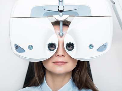
Digital refraction with a phoropter.
This is typically the main reason patients come for a yearly check up as this test is used to determine the eyeglasses prescription. All of our exam lanes are equipped with the most advanced technology and we will never make you feel like you’re being rushed.
Autorefractor and Keratometer
An autorefractor is a quick test that gives an estimate of the eyeglass prescription. It is helpful as a starting point that will then be fine tuned. A keratometer measures the curvatures of the cornea. This is helpful in detecting various corneal diseases as well as determining which contact lens would fit the patient the best and improve overall comfortability.
Slit-lamp exam
A slit lamp is a binocular microscope (or biomicroscope) used to examine the structures of the eye under high magnification, including eyelids, cornea, conjunctiva, iris, and lens. With the help of a hand-held lens, the doctor may also use the slit lamp to examine structures located farther back in the eye, such as the retina and optic nerve.
The slit lamp exam can be used to detect a wide range of eye conditions and diseases, including cataracts, macular degeneration, corneal ulcers, and diabetic retinopathy, etc.
The Glaucoma Test
Similar to blood pressure, ocular pressure is most often asymptomatic. Most people remember (and fear) the dreadful puff of air to check intraocular pressure. We will NEVER use that outdated test. Our top of the line tonometer is completely painless so NO AIR PUFF!
Visual Field Test
This test checks for the possible presence of blind spots (scotomas) in the peripheral (side) vision by performing a visual field test. These blind spots can originate from eye diseases like glaucoma or may help identify specific areas of brain damage caused by a stroke or tumor.
Dilation versus Optomap
Dilation involves putting eyedrops in to enlarge the pupil so we can check the health of the retina in the back of the eye. Typically, most patients can drive home and side effect of light sensitivity and mild blurred vision last anywhere from 3-6 hours
Optomap is encouraged on all patients and provides an ultrawide field photograph of the retina WITHOUT DILATION. As an added bonus, we get to monitor any changes in appearance and compare year after year.
How long will the appointment take?
A comprehensive eye exam can take 1 hour or longer, depending on the number and complexity of tests required to fully evaluate vision and the eyes health.
Can I drive after my eyes are dilated?
Many patients are able to drive themselves after having their eyes dilated, but it is important to remember that eyes will be sensitive to light and vision may be blurry and you should wear sunglasses after your exam. With Optomap, we can avoid dilation all together by using Optomap for retinal imaging.
Quick Links
Helpful Articles
article_category: ocular disease management
article_category: exotic
article_category: restorative
article_category: products
article_category: general
article_category: preventative
article_category: Aesthetics
article_category: contact lenses
article_category: health
article_category: cats
article_category: services
article_category: large animal
article_category: surgical procedures
article_category: psychiatry
article_category: conditions
article_category: cosmetic
article_category: eye surgery co-management
article_category: dogs
article_category: technology
article_category: FAQ & tips
article_category: eye health
article_category: eyeglasses
[]
Flowbite is an open-source library of interactive components built on top of Tailwind CSS including buttons, dropdowns, modals, navbars, and more.
Check out this guide to learn how to get started and start developing websites even faster with components on top of Tailwind CSS.
All Eyecare Services
We offer a wide variety of eye care services to the Madison community. Contact us with any questions about our services.

Keep In Touch
For non-urgent questions or to learn more about our services, contact us today!
© 2025 North Coast Optical . All rights Reserved - Accessibility Statement - Privacy Policy - Sitemap
Managed and Designed by
![]()